
Mammalian cell display services
When your target demands native glycosylation, disulfide bonding, or complex multi-subunit assembly, mammalian display provides the right biological context for screening
Discuss your project →Screening in a native human cellular context
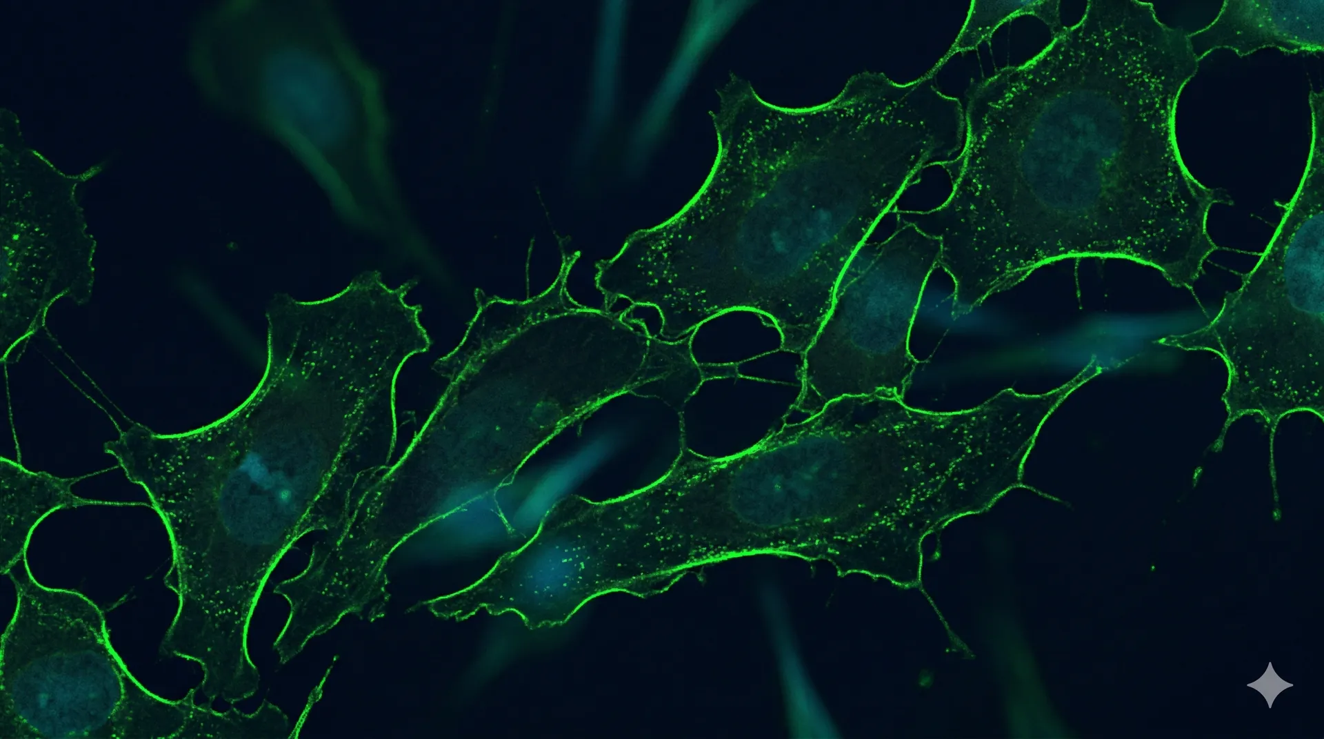
Mammalian display screens protein libraries on HEK293 cells, providing human-pattern N-glycosylation, native disulfide architectures, and proper membrane embedding. Hits selected in this context carry post-translational modifications that directly reflect the final production format.
Library delivery uses lentiviral transduction or Cas9-mediated integration to ensure single-copy payload per cell. This one-cell-one-variant linkage enables quantitative FACS-based selection without avidity artifacts from multi-copy expression. Full-length IgG antibodies, multi-pass transmembrane proteins, and targets with glycan-dependent epitopes all benefit from mammalian display.
Targets that require mammalian cell display
Glycan-dependent epitopes
Targets where the binding epitope includes or is conformationally dependent on N-linked glycans. Yeast hyperglycosylation produces mannose-rich structures that differ from human glycoforms, potentially masking or altering critical epitopes.
Full-length IgG screening
Display of complete IgG1 or IgG4 antibody formats that require proper heavy-light chain pairing, glycosylation of the Fc domain, and native disulfide bond formation. Avoids reformatting artifacts seen when converting scFv hits to full-length.
Multi-pass transmembrane targets
GPCRs, ion channels, and other multi-pass membrane proteins that must be presented in a native lipid bilayer context. Mammalian cells provide the correct membrane composition and chaperone machinery for proper folding and surface trafficking.
Developability-focused campaigns
When downstream manufacturing will use CHO or HEK production, screening in the same cellular context reduces the gap between discovery and development. Hits selected in mammalian display are more likely to express well in production cell lines.
Single-copy integration for quantitative screening
Low-MOI viral integration
Lentiviral delivery at low multiplicity of infection ensures single-copy integration per cell. Stable genomic insertion provides consistent expression across cell divisions, enabling multi-round selection without library loss.
- • Stable integration across passages
- • Established protocol for large libraries
- • Compatible with extended selection campaigns
Site-specific genomic insertion
CRISPR/Cas9-directed integration at a defined safe harbor locus. Every cell expresses its payload from the same genomic context, eliminating position-effect variation and enabling direct comparison of expression levels across variants.
- • Uniform expression from defined locus
- • No position-effect variability
- • Quantitative expression-level comparisons
Mammalian display screening workflow
Library construction
Gene synthesis of your diversified library, cloned into mammalian expression vectors with surface display tags. Quality-controlled by NGS before transfection.
Transfection and display
Electroporation or lipofection into CHO or HEK293 cells. Surface expression confirmed by flow cytometry before selection begins.
FACS selection
Multi-round fluorescence-activated sorting against labeled antigen. Increasing stringency each round. Counter-selection against off-targets as needed.
NGS and hit calling
Enriched populations sequenced and analyzed. Quantitative enrichment ratios identify top candidates. Ranked hit lists delivered with enrichment metrics.
Mammalian display vs yeast display
Most campaigns start on yeast display for diversity and speed, then move to mammalian display when the target or the final format demands a human cellular context. Here is how the two compare and where each one wins.
Diversity, speed, and quantitative FACS
Libraries to 10^8-10^9, fast cycle times, and a per-clone affinity readout by flow cytometry. The default platform for nanobody and scFv discovery and affinity maturation. Installs only high-mannose glycans, so glycan-dependent epitopes may not be presented natively.
Native PTMs and full-length format
Human glycosylation, full-length IgG assembly, native disulfide architecture, and multi-pass membrane context. Lower diversity (10^5-10^6), but hits reflect the final production format and any glycan-dependent epitopes.
The standard workflow uses both: discover and mature on yeast display, then validate developability and glycan-dependent binding on mammalian display. See the two-platform approach for costs and timeline.
Technical articles on mammalian display
When to choose mammalian over yeast display
A decision guide for the platform call: glycan-dependent epitopes, full-length IgG, membrane targets, and production-context developability.
Full-length IgG display on mammalian cells
Screening complete IgG with native heavy-light pairing and Fc glycosylation, and why it beats reformatting scFv hits.
Mammalian cell display: when CHO and HEK293 outperform yeast
The targets where human-like glycosylation and full-length IgG context justify the slower, costlier platform.
Glycoprotein engineering: yeast vs mammalian
Why glycan-dependent epitopes and effector function force the screen onto mammalian cells.
Yeast and mammalian display: two-platform approach
Affinity discovery on yeast, developability validation on mammalian, with costs and timeline.
PTMs in mammalian protein production
How glycosylation and other modifications shape developability and the data you can trust.
Mammalian cell engineering
From stable line to functional assay, the host-cell work that display campaigns sit on top of.
Troubleshooting low display levels
A step-by-step checklist for surface-expression failures on yeast and mammalian cells.










Foundational papers in mammalian display
The peer-reviewed methods behind mammalian cell display for antibody discovery and maturation.
Mammalian display questions
When should I use mammalian display instead of yeast display? +
Choose mammalian display when the epitope is glycan-dependent, when you need full-length IgG with native Fc glycosylation, when the target is a multi-pass membrane protein, or when downstream manufacturing in CHO or HEK makes production-context developability part of the selection criteria.
Do you display full-length IgG? +
Yes. Mammalian display presents complete IgG1 or IgG4 with proper heavy-light chain pairing, Fc glycosylation, and native disulfide formation, which avoids the reformatting artifacts seen when scFv hits are later converted to full-length antibodies.
CHO or HEK293 — which cell line do you use? +
HEK293 is the default for fast transfection and human-pattern glycosylation. We use CHO when the goal is to match a CHO production line so that surface-display hits carry over to manufacturing with minimal expression surprises.
What library sizes does mammalian display support? +
Roughly 10^5 to 10^6 per integration — lower than yeast display, but single-copy genomic integration gives clean, quantitative one-cell-one-variant readout without avidity artifacts.
Can you discover antibodies directly on mammalian display, or only validate? +
Both. We run de novo discovery against complex targets that yeast cannot present natively, and we use mammalian display to validate developability and glycan-dependent binding of hits first found on yeast or phage.
How does mammalian display fit with yeast display? +
In the two-platform approach: yeast display delivers diversity, speed, and affinity ranking, then mammalian display confirms the leads in a human cellular context and in the final antibody format. Many campaigns use both in sequence.
Need mammalian-context screening?
Describe your target and the post-translational requirements. We will recommend the optimal display platform and return a detailed proposal.
Start a project →