Receptor-targeting binder design
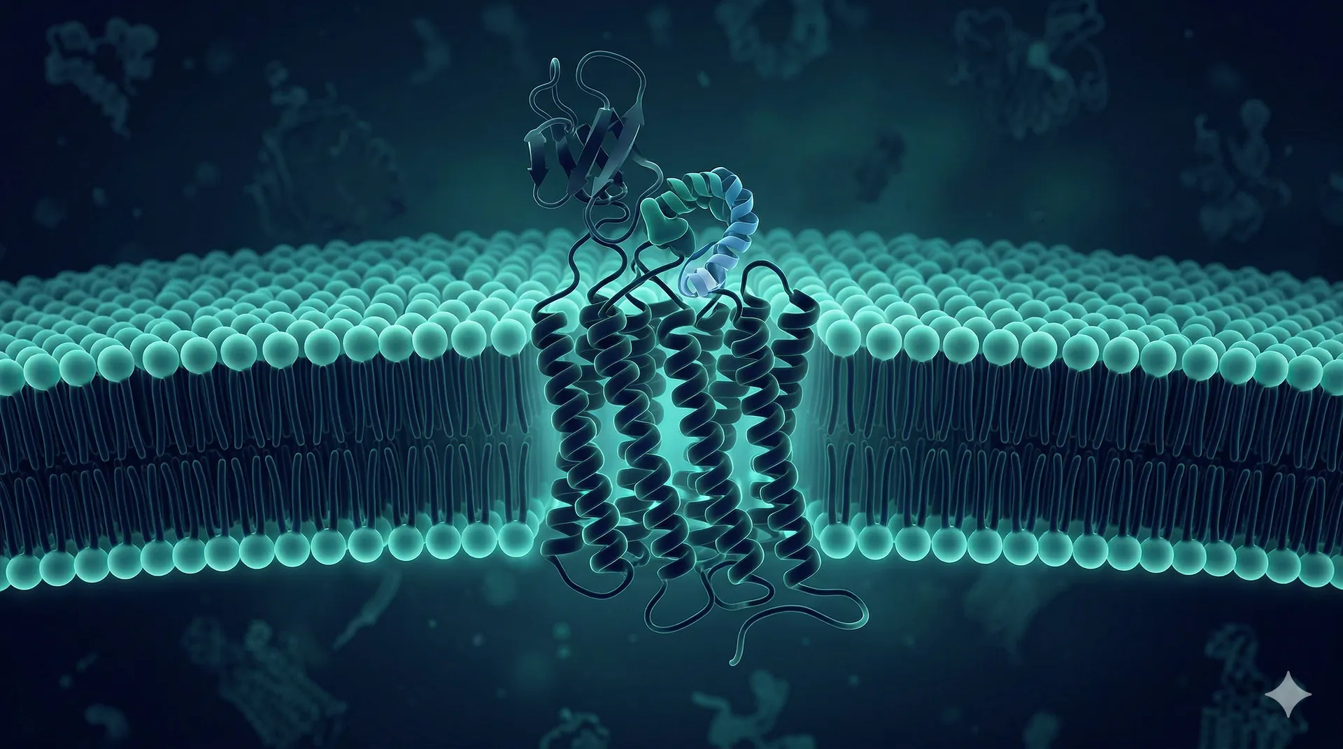
GPCRs, ion channels, and receptor tyrosine kinases are among the most valuable and most difficult drug targets. De novo design offers a structural approach where conventional methods stall.
Discuss your target →Why membrane receptors resist conventional binder discovery
Membrane proteins present a unique set of challenges for binder discovery. GPCRs expose small extracellular loops with limited surface area for binding. Ion channels present conformationally dynamic epitopes that shift between open, closed, and inactivated states. Receptor tyrosine kinases form transient dimeric interfaces that are difficult to target with traditional approaches.
Conventional antibody discovery methods — immunization and phage display — struggle with these targets because the proteins are difficult to express as soluble antigens, their native conformations depend on the lipid bilayer, and the epitopes accessible in solution differ from those on the cell surface.
De novo computational design addresses these challenges by working directly from the target structure, designing binders that engage specific surface patches regardless of their size, flatness, or conformational flexibility.
Small extracellular epitopes
GPCRs expose limited surface area outside the membrane. De novo design can target patches as small as 500-800 square angstroms.
Conformation dependence
Ion channel states change epitope accessibility. Design can target specific conformational states using state-locked structures.
Membrane context required
Native conformation depends on the lipid bilayer. Mammalian display presents targets in their native membrane environment.
Glycan shielding
Heavy N-glycosylation masks receptor surfaces. Computational design can thread binders through glycan-free windows.
GPCR, ion channel, and RTK binder design
GPCR binders
G protein-coupled receptors are the largest family of drug targets, yet most GPCR drugs are small molecules. Protein binders targeting extracellular loops and N-terminal domains offer allosteric modulation, receptor stabilization, and conformation-selective targeting that small molecules cannot achieve.
Ion channel modulators
Binders targeting the extracellular vestibule, turret, or pore-loop regions of voltage-gated and ligand-gated ion channels. Applications include pain (Nav, TRPV), cardiac (hERG), and neurological (GABA, NMDA) targets where protein binders offer subtype selectivity.
Receptor tyrosine kinase binders
Targeting the extracellular domains of EGFR, HER2, VEGFR, FGFR, and other RTK family members. De novo binders can engage epitopes distinct from those targeted by existing therapeutic antibodies, accessing novel mechanisms of action.
Cytokine receptor binders
Binders for interleukin receptors, TNF receptor superfamily members, and other single-pass transmembrane receptors. Blocking ligand binding, stabilizing inactive conformations, or engaging allosteric sites on the extracellular domain.
Immune checkpoint receptors
Novel binders for PD-1, CTLA-4, LAG-3, TIGIT, and emerging checkpoint targets. De novo design provides access to non-competitive epitopes and novel binding geometries that may produce differentiated pharmacology.
Orphan receptors
Receptors with no known natural ligand and limited existing tool compounds. De novo design requires only a structural model, making it one of the few viable approaches for generating binding reagents against orphan targets.
Design-to-validation workflow for membrane protein targets
Membrane protein targets require adaptations to the standard binder discovery workflow — particularly in how the target is presented during screening.
Structure-based design
RFdiffusion generates binder backbones conditioned on accessible extracellular epitopes identified from cryo-EM or AlphaFold structures. Designs are filtered for predicted binding energy and absence of membrane-clashing geometry.
Cell-surface target presentation
Instead of soluble antigen, the target receptor is overexpressed on cell surfaces in its native membrane context. Binder library and target-expressing cells are co-incubated for cell-based selection.
Mammalian display screening
Binder library displayed on mammalian cells, selected against target-expressing cells by FACS. Alternating positive selection (target-high cells) and negative selection (target-null cells) ensures specificity.
Functional characterization
Confirmed binders tested for receptor modulation: agonism, antagonism, allosteric effects, or neutral binding. Cell-based functional assays (calcium flux, cAMP, phosphorylation) alongside binding confirmation.
Where de novo design excels for receptor targets
Computational protein design provides specific advantages for membrane receptor targets that are difficult or impossible to replicate with traditional binder discovery approaches.
Epitope precision
Specify exact hotspot residues on the receptor extracellular domain. Design binders that engage a specific loop, a subunit interface, or a conformational epitope that is only accessible in a particular receptor state. This level of epitope control is not achievable with immunization-based methods.
No soluble antigen required
Immunization and phage display require soluble, purified target protein. Many membrane receptors cannot be produced as stable soluble ectodomains. De novo design works from the structure alone, and screening uses cell-surface-presented receptor in its native membrane context.
Conformation-selective targeting
Design binders against specific receptor conformational states — active, inactive, ligand-bound, or apo. Use state-locked structures (disulfide-stabilized, ligand-trapped) as design templates to generate conformation-selective binders with built-in pharmacological specificity.
Subtype selectivity by design
Closely related receptor subtypes (e.g., Nav1.7 vs. Nav1.5, FGFR1 vs. FGFR2) share high sequence identity in their druggable domains. Computational design can exploit subtle structural differences between subtypes to generate selective binders from the outset.
Targeting a difficult receptor?
Share your receptor target, the available structural data, and the desired mechanism of action. We will assess feasibility and propose a de novo binder design campaign.
Start a project →